3D Cell Culture Imaging & Reconstruction Platform
At Tissuetinker, we needed a better understanding of the 3D architecture and viability of our tumoroid cultures, so I developed a custom data and image processing platform. Without having access to any confocal microscopes, I designed the platform to reconstruct a 3D environment from a collection of 2D images taken at different depths (z-stacks).
It functions by first aligning all of the images, then creating a 3D rendition from the images, and finally isolating any objects within based on relative signal intensities and sharpness. We can then choose to isolate detected tumoroids/cells/debris by size exclusion to get a more accurate reading of cell and tumoroid viability and sizes.
Coded in Python utilizing the napari image viewing framework
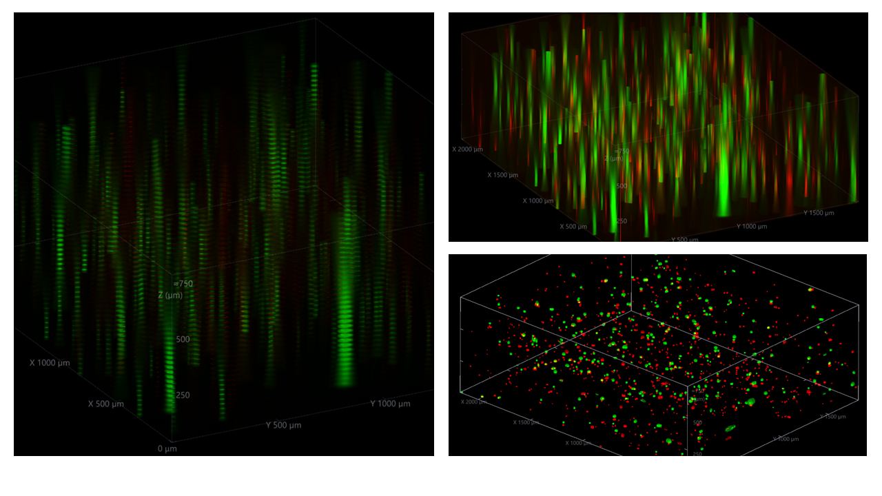
Images obtained from an ECHO Revolve immunofluorescent microscope using a Calcein AM + Ethidium homodimer-1 (EthD-1) assay for viability.